
The Hidden Scar on Your Lungs
By March 2026, we are three years past the initial peak of the pandemic, but for many people, the virus has not fully let go. You might feel fine most of the time, yet still struggle to catch your breath when climbing stairs or carrying groceries. This is often called pulmonary long COVID, defined as a set of persistent respiratory symptoms that continue months after the initial infection has cleared. It affects roughly one-third of all long COVID cases, according to research from the Centre for Heart Lung Innovation published in 2025.
This isn't just about feeling tired. Research led by Dr. Don Sin at the University of British Columbia identified that immune cells keep attacking the lungs even after the virus is gone. It's a silent battle happening deep inside the smallest airways. Many patients walk away from the hospital thinking they have recovered, only to find their fitness levels haven't returned to normal a year later.
What Is Actually Happening Inside?
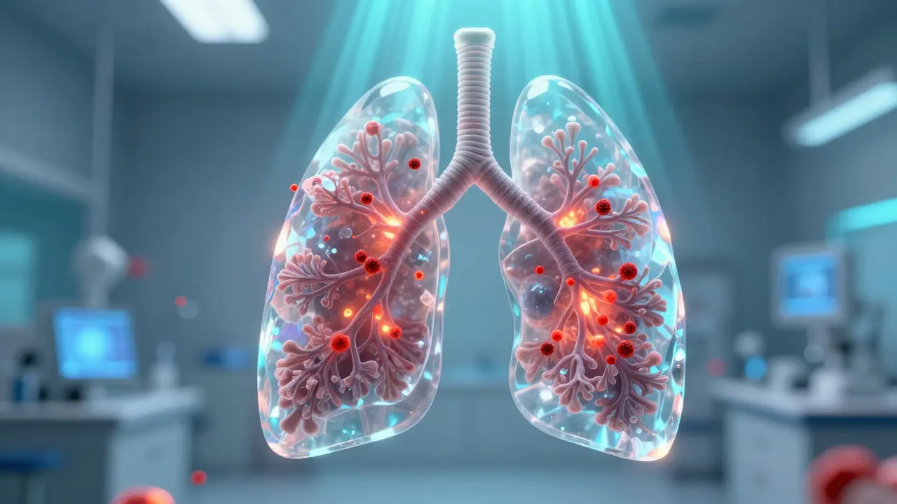
To understand why you still feel winded, we have to look at what happens during recovery. When you contract SARS-CoV-2, your immune system sends out soldiers called neutrophils to fight the infection. Normally, these cells do their job and then leave. In people with pulmonary long COVID, these cells stay behind. Dr. Sin described them as "dirty bombs," continuing to trigger inflammatory responses even though the virus is no longer present.
This persistent inflammation damages the small airways in ways we used to miss. Standard lung scans often come back looking "normal," which leaves patients confused when they still can't run up a flight of steps. However, newer technology is changing how doctors see this. Hyperpolarized xenon MRI technology allows us to visualize oxygen transfer in the tiny parts of the lung where traditional X-rays and CT scans fail. Studies from 2024 and 2025 showed distinct clusters of gas exchange abnormalities using this method, confirming that damage exists even if function tests appear normal.
Can Standard Tests Detect the Problem?
If you visit your doctor complaining of shortness of breath, they will likely order a spirometry test to measure your lung capacity. While useful, this test often misses the subtle damage caused by long-term inflammation. The HLI biobank research revealed that single-cell sequencing is needed to spot the persistent neutrophilic inflammation mentioned earlier.
In 2025, the von LL O'Mahoney systematic review analyzed 50 studies involving thousands of patients. It found that SARS-CoV-2 infection increases the risk of long-term respiratory symptoms by 2.6-fold, regardless of whether you were hospitalized initially. However, the severity matters. Patients who spent time in the hospital are more likely to develop post-COVID-19 pulmonary fibrosis (PCPF), where scar tissue forms and hardens lung tissue permanently.
| Diagnostic Tool | Detects Small Airway Damage | Best Used For |
|---|---|---|
| Spirometry (Lung Function Test) | Limited | Measuring airflow obstruction and capacity |
| Chest CT Scan | Moderate | Spotting large scarring or obvious structural changes |
| Hyperpolarized Xenon MRI | High | Visualizing gas exchange in smallest airways |
| 30STS Test (Sit-to-Stand) | N/A (Functional) | Assessing physical capacity and fatigue impact |
Risks: Who Needs Extra Care?
Not everyone faces the same level of risk. A Korean retrospective study covering 688 patients documented that 12.6% of hospitalized individuals developed PCPF. If you had mild symptoms at home, your risk drops significantly. However, certain groups face higher stakes. People with pre-existing conditions like chronic obstructive pulmonary disease (COPD) saw mortality rates jump to 4.6% compared to 0% in non-COVID patients.
Another critical factor is your functional baseline before infection. The RECOVER Initiative's update in September 2025 noted that those already experiencing burdensome fatigue performed significantly fewer repetitions on sit-to-stand tests, indicating lower reserves to handle the viral assault. If you were already struggling with heart failure or diabetes, the toll on your lungs was often compounded.
Interestingly, the treatments you received during the acute phase play a role. That same Korean study observed that remdesivir use correlated with a reduced risk of PCPF development. Conversely, baricitinib usage was linked to a slightly increased risk, though researchers noted this could be due to bias in who received the drug rather than a direct cause. Always discuss medication history with your specialist to rule out other causes.
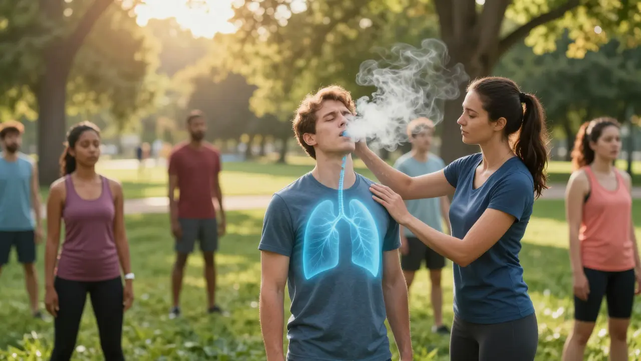
Rehabilitation: Rebuilding Your Breath
Once the dust settles, what can you actually do about it? Pulmonary rehabilitation remains the gold standard for recovery. Unlike simple rest, these programs challenge your body to adapt without burning out. A typical program runs for 8 to 12 weeks, requiring sessions two to three times weekly. It starts after the acute phase, usually four weeks post-infection, once stable vitals are confirmed.
You won't just be running on a treadmill. Protocols focus on specific breathing exercises designed to clear mucus and retrain the muscles involved in respiration. The Lung Foundation Australia reviewed multidisciplinary approaches showing improvements in Forced Expiratory Volume (FEV1) and diffusion capacity. One key metric they tracked was the 6-minute walk distance (6MWD), which consistently improved alongside reduced reports of dyspnea (shortness of breath).
For those with significant scarring, pacing is vital. The RECOVER Initiative highlighted that patients with very burdensome symptoms, including post-exertional malaise (PEM), must monitor their activity closely. Pushing through pain often leads to crashes that undo weeks of progress. Instead, aim for steady, small increments in activity duration rather than intensity spikes.
Tracking Your Recovery Milestones
How do you know if the rehab is working? You need objective numbers, not just feelings. Clinicians often use the mMRC Dyspnea Scale to track your breathlessness scores over time. If your score stays above 2 at the one-month mark, you are statistically more likely to have persistent dysfunction requiring intensive support.
Another practical tool is the 30STS test. This measures how many times you can stand up from a chair without arms in 30 seconds. It's simple, requires no expensive equipment, and provides a clear benchmark for functional improvement. Data from 2025 suggests that measurable functional gains correlate strongly with better quality of life outcomes.
We also look at blood tests and imaging to ensure inflammation markers are dropping. While xenon MRI is becoming more common, standard CT scans taken six months apart can still show trends in fibrosis progression. Ideally, you want stability or slight improvement; rapid worsening signals a need to adjust your medical regimen immediately.
Looking Ahead: What Does 2026 Hold?
As we move through 2026, the landscape of treatment is shifting from pure management to targeted therapy. The HLI team is expanding their network to track treatment responses specifically for those with small airway inflammation. Early trials suggest that targeting the neutrophil activity directly could halt the cycle of damage.
The American Thoracic Society and European Respiratory Society are currently finalizing specialized guidelines for these complications, anticipated for release late in 2025 and updated through 2026. This means clearer criteria for diagnosis and treatment plans for primary care physicians. Furthermore, industry analysts predict a boom in accessible pulmonary rehabilitation tools designed specifically for the post-viral population, making professional-grade recovery possible at home under guidance.
While the road to full recovery varies for everyone, the outlook remains cautiously optimistic. Evidence shows that most patients see measurable lung function improvements within six months of dedicated rehabilitation. Permanent fibrotic changes affect a smaller subset of the population, specifically those who endured severe illness requiring ventilation. For the majority, consistent effort combined with medical monitoring yields tangible results.
How long does lung recovery take after COVID-19?
Most patients experience gradual improvements in lung function over a period of six months. However, some symptoms may persist beyond a year if neutrophilic inflammation continues. Active rehabilitation programs typically run for 8 to 12 weeks to maximize recovery speed.
Will a chest X-ray detect lung damage?
Standard chest X-rays often miss damage in the smallest airways. Advanced imaging like hyperpolarized xenon MRI is better suited to reveal gas exchange problems. If you have breathing issues despite a normal X-ray, ask your doctor about referral for specialized functional testing.
What is the best exercise for long COVID lung patients?
Structured pulmonary rehabilitation is superior to unguided exercise. This includes aerobic conditioning combined with specific breathing exercises. Start with low-intensity activities like walking and progress based on your 6-minute walk distance capability to avoid overexertion.
Does having COPD increase my risk?
Yes, existing COPD significantly increases the risk of severe outcomes. Studies show a 4.6% mortality rate for COPD patients contracting COVID-19 versus zero for non-COVID cases. Close monitoring for exacerbations is essential during recovery.
Can inflammation be treated years later?
Research in 2025 and 2026 is exploring anti-inflammatory therapies targeting persistent neutrophil activity. While guidelines are being finalized, current management focuses on stabilizing symptoms through physical therapy and avoiding secondary infections to prevent further scarring.


15 Comments
Rohan Kumar
Another study says something we already know lol 😒🤔
Sabrina Herciu
The data regarding neutrophil persistence is extremely significant! The implications for treatment protocols are massive! Hyperpolarized xenon MRI changes everything we knew about scanning! Dr. Sin's team should be celebrated for this breakthrough! Research from 2025 confirms what patients felt deep inside! We need wider access to these specialized tools immediately!!!
Monique Ball
It is really wonderful to see all these new technologies emerging for our recovery journey today! We often feel like the medical world has forgotten us completely during the darkest days. But studies like the von LL O'Mahoney review give us such a massive boost of confidence regarding prognosis!! 🌟 Everyone here should know that even small improvements mean progress toward a better life. The rehabilitation programs mentioned are absolutely gold for rebuilding lung capacity slowly over time! 🏃♀️ You cannot rush healing and you must listen to your own body limits very carefully!! 🛑 Ignoring post-exertional malaise is a terrible mistake that many of us learn the hard way unfortunately! 😢 We need to track our six-minute walk distance consistently every single month! 📏 It takes courage to push through the fatigue barriers that hold us back mentally! 💪 Family support systems are critical for maintaining motivation throughout the twelve-week process! 💕 Doctors are finally listening to the subtle symptoms that were ignored before this pandemic started! 🩺 The hyperpolarized xenon MRI technology sounds like a miracle for diagnosis accuracy rates! ✨ Please remember that inflammation markers take months to stabilize in blood work results! 🧬 Stay positive because the outlook for the majority of patients remains cautiously optimistic according to experts! 📈 We will get through this health crisis together one breath at a time definitely!! ❤️
gina macabuhay
Your naive enthusiasm ignores the grim statistical realities facing fibrosis patients right now. Many individuals permanently lose function regardless of their optimistic mindset or therapy regimen. You ought to stop spreading misinformation disguised as encouragement to vulnerable survivors.
Austin Oguche
Discipline remains the cornerstone of effective pulmonary rehabilitation protocols globally
Debra Brigman
The breath is the tether between soul and matter yet we fight for it with clinical precision instead of poetic reverence for survival
tyler lamarre
Most people lack the IQ to follow complex rehab plans anyway so they blame the virus instead of their own failure. It is amusing how little understanding exists outside the upper class hospitals.
Tony Yorke
Consistency wins over intelligence every single time in recovery
Poppy Jackson
My heart breaks for those stuck in hospital beds still breathing through tubes and hoping for tomorrow please hold onto hope my friends
Jeannette Kwiatkowski Kwiatkowski
Stop feeling sorry for yourself and just go lift weights because weakness breeds more weakness in the body. It is simple biology not magic spells.
Aaron Olney
i havent been able to run since feb and doctors just shrug shoulders at me its unfair why does this hapen to me when i did everything right
Sophie Hallam
Frustration is natural but regional availability of care determines the options available to you currently. Medical testing varies significantly by location.
Philip Wynkoop
Staying strong 💪❤️
Richard Kubíček
Progress is slow but real for everyone who puts in the work. The future holds promise if we stay collaborative and respectful of our individual journeys forward.
kendra 0712
We need more research funding NOW!!!!! The guidelines coming in 2026 will change everything for primary care physicians!!! Keep pushing for better diagnostics everywhere!